АвтоАвтоматизацияАрхитектураАстрономияАудитБиологияБухгалтерияВоенное делоГенетикаГеографияГеологияГосударствоДомДругоеЖурналистика и СМИИзобретательствоИностранные языкиИнформатикаИскусствоИсторияКомпьютерыКулинарияКультураЛексикологияЛитератураЛогикаМаркетингМатематикаМашиностроениеМедицинаМенеджментМеталлы и СваркаМеханикаМузыкаНаселениеОбразованиеОхрана безопасности жизниОхрана ТрудаПедагогикаПолитикаПравоПриборостроениеПрограммированиеПроизводствоПромышленностьПсихологияРадиоРегилияСвязьСоциологияСпортСтандартизацияСтроительствоТехнологииТорговляТуризмФизикаФизиологияФилософияФинансыХимияХозяйствоЦеннообразованиеЧерчениеЭкологияЭконометрикаЭкономикаЭлектроникаЮриспунденкция
Phase - contrast method
|
Читайте также: |

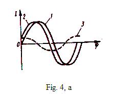
The method is applied for supervision of non contrast objects; it is based on use of a difference of phases which is formed at passage of light through various structures of investigated object. Intensity of the light wave, which are passing through transparent object, does not change almost, but phases change. These changes depend upon thickness of object and its parameter of refraction. Transparent objects is called out-phasing. Thus to see details of such objects it is impossible. In biophysical researches such objects are necessary for painting, however thus their properties and viability can change. The transparent environment is established on a way of a light bunch. This environment there is a transparent inclusion, for example, a bacterium (fig. 3). The passing bunch of light will be divided on two parts, the first part will pass through transparent object and by a lens will be focused on site Ф of focal plane F (see fig. 4 a, a line 1).
Other part will do diffraction on heterogeneity of object and will gather by lens in a point A of plane I (see fig. 4 b, a line 2). The curve 3 grows out diffraction of light on a bacterium. The difference of phases of curves arises because of different parameters of refraction of environments.

The eye in plane I does not distinguish a wave 1 and 2 as their intensity identical, and eyes does not react on distinction of phases. It is necessary to transform a phase difference in amplitude difference. For this purpose in plane F it is necessary to put the small round phase plate absorbing a wave 1, in this case contrast of a bacterium it will be strengthened.
Phase-contrast devices (a plate, sate of lenses) are additional devices to a microscope.
Поиск по сайту: