АвтоАвтоматизацияАрхитектураАстрономияАудитБиологияБухгалтерияВоенное делоГенетикаГеографияГеологияГосударствоДомДругоеЖурналистика и СМИИзобретательствоИностранные языкиИнформатикаИскусствоИсторияКомпьютерыКулинарияКультураЛексикологияЛитератураЛогикаМаркетингМатематикаМашиностроениеМедицинаМенеджментМеталлы и СваркаМеханикаМузыкаНаселениеОбразованиеОхрана безопасности жизниОхрана ТрудаПедагогикаПолитикаПравоПриборостроениеПрограммированиеПроизводствоПромышленностьПсихологияРадиоРегилияСвязьСоциологияСпортСтандартизацияСтроительствоТехнологииТорговляТуризмФизикаФизиологияФилософияФинансыХимияХозяйствоЦеннообразованиеЧерчениеЭкологияЭконометрикаЭкономикаЭлектроникаЮриспунденкция
Structure of a cross-section-striped muscle. Model of sliding strands
The muscular tissue is set of muscular cells (fibres), extracellular substance (collagen, elastin, etc.) and a dense network of nerve fibre and blood vessels. Muscles can be divided by structure on to two groups: smooth - muscles of bowels, a wall of vessels, and cross-section-striped - skeletal, muscles of heart. Independentlyfrom a structure all of them have similar mechanical properties, the identical mechanism of activation and similar chemical composition.
The cross-section-striped structure of muscular fibres can be observed under optical microscope. The separate muscular fibre has diameter 20 - 80 microns and is surrounded by a plasmatic membrane with thickness 10 nanometers. Each separate fibre is strongly elongated cell. The length of separate fibres {cells) can essentially vary depending on a kind of a muscle from hundreds micron up to several centimeters. Inside of a fibre, except for known organelles (a nucleus, nucleolus, mitochondrion, Golgi apparatus, etc.), there are contractive apparatus of the cells, consisting of 1000 - 2000 in parallel located myofibrillas with diameter 1-2 microns, and also cellular organelles: sarcoplasmic reticulum and system of cross-section tubules - T-system.
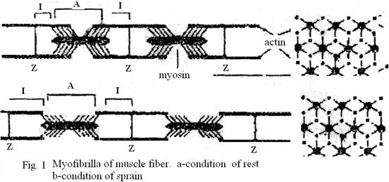
Myofibrilla (Fig.1) has 2 zones: A -zone - dark line which in polarized light give double refraction, i.e. possess property of anisotropy (from here the name: A-zone), I-zone - the light lines, not giving double refraction, that is isotropic (from here the name: I-zone). In the field of an I-zone there is a dark narrow line - Z-disk. The interval between two Z-disks is called sarcomere, it is elementary contractive unit of a muscular cell.
 Sarcomere is the ordered system of the thick and thin strands, located hexagonal in cross-section section. Thick strand has thickness - 12 nanometers and length - 1,5 microns and consists of fiber of myosin. The thin strand has diameter of 8 nanometers, length 1 micron and consists of fiber actin, attached by one end to a Z-disk.
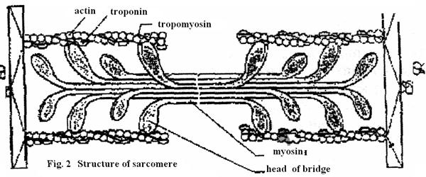
Actin strand consists of two twirled one around of other monomers of actin with thickness on 5 nanometers (fig. 2). This structure is similar to two strands of a beads, braided on 14 beads in a coil. Molecule of troponin are built to circuits of actin regular approximately through 40 nanometers, and the circuit covers a strand of tropomyosin. At reduction of a muscle thin strands are moved between thick strands. There is a relative sliding of strands without change their lengths. This process is caused by interaction special ledges of myiosin - cross-section ponticulus with the active centers located on actin. Ponticulus depart from thick strands periodically on distance of 14,5 nanometers from each other.
In the weakened condition of myofibrillas of molecule of tropomyosin block an attachment of cross-section ponticulus to actin circuits (Fig.3.а).
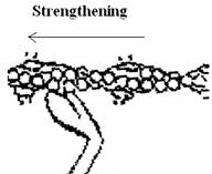


Fig. 3. A process of activating of ponticulus and generation of strngthening in a sarcomere.
Ions of Ca activate ponticulus and open the areas of their attaching to aktin (fig.3). As a result the ponticulus of miosin register to the aktin strands, molecules fission ATP and confirmation of ponticulus changes: their heads turn into a sarcomere (fig.3).
It leads to the generation of force, to sliding of aktin relatively to the thick strands of miosin to the center of sarcomere, that causes shortening of muscle. After completion of activation ponticulus is opened and a sarcomere goes back into the initial condition. At shortening the volume of sarcomere does not change, consequently, he becomes thicker, what is confirmed on the pictures of cross-sectional of muscles by an electronic microscopy. Every cycle opening-closing is accompani ed breaking up to one molecule of ATP.
Thus, actin-miosin a complex is the chemical converter of energy АТP. The considered structure and sequence of processes is called model of sliding strands.
For the first time sliding of strings in sarcomere was revealed English scientist H.Haksli. It has formulated model of sliding strings.
Experimental data about a microstructure of muscles have been studied by means of electronic microscopy, X-rays analysis.
Basic provisions of model of sliding strings:
1. Lengths of strands of actin and miosin during reduction do not change.
2. Change of length of sarcomere at reduction - result of relative longitudinal displacement of strands of actin and miosin.
3. The cross-section ponticulus departing from miosin, can connect to complementary centers actin.
4. Ponticuluses are attached to actin not simultaneously.
5. The closed bridges develop effort, then they open.
6. Reduction and a relaxation of a muscle consist in increase and the subsequent reduction of number of the bridges making a cycle closing - opening.
 7. Each cycle is connected with hydrolysis of one molecule of АТP.
8. Acts of closing - opening of ponticuluses occur independently from each other.
 Fig. 4. Dependence of the maximal value of developed force on a degree of overlapping actin and miosin strings.
Opportunity of sarcomere to be reduced and develop effort depends on entry conditions. If sarcomere will stretch (its length 3,65 microns) bridges are not blocked by actin strings and at stimulation of such element the effort is not formed (1 on fig. 4 a)
If sarcomere is in a working initial condition (the size of sarcomere - 2,2 microns) at stimulation it will develop the maximal force (2 on fig. 4 a). If the initial size of sarcomere too small, generation of effortn decreases (an arrow and a fragment 5).
Поиск по сайту: