 |
АвтоАвтоматизацияАрхитектураАстрономияАудитБиологияБухгалтерияВоенное делоГенетикаГеографияГеологияГосударствоДомДругоеЖурналистика и СМИИзобретательствоИностранные языкиИнформатикаИскусствоИсторияКомпьютерыКулинарияКультураЛексикологияЛитератураЛогикаМаркетингМатематикаМашиностроениеМедицинаМенеджментМеталлы и СваркаМеханикаМузыкаНаселениеОбразованиеОхрана безопасности жизниОхрана ТрудаПедагогикаПолитикаПравоПриборостроениеПрограммированиеПроизводствоПромышленностьПсихологияРадиоРегилияСвязьСоциологияСпортСтандартизацияСтроительствоТехнологииТорговляТуризмФизикаФизиологияФилософияФинансыХимияХозяйствоЦеннообразованиеЧерчениеЭкологияЭконометрикаЭкономикаЭлектроникаЮриспунденкция
Computer tomograph
|
Читайте также: |
 The computer tomograph (CT) is a combination of x-ray installation and a computer. X-ray installation does pictures of the patient under different corners, (cuts) which are processed and summarized by a computer - the image allowing doctors "to glance" inside of a body of the patient turns out.
Computer tomography it is used now in increasing frequency. This method nonivasive (does not demand operative intervention), safe and it is applied at many diseases. CТ ideally approaches for diagnosing bone damages and traumas. Besides the fresh bleeding is well visible on CТ, therefore CТ is applied at researches of patients with traumas of a head, a thorax and belly and pelvic cavities, and also insults in an early (!) stage. Use of contrast substance allows receiving the qualitative image of vessels, kidneys and intestines. By means of a computer tomography it is possible to investigate practically any body - from a brain up to bones. Often computer tomography is used for specification of the pathologies revealed by other methods. For example, at an antritis, all over again rontgenography of additional bosoms of a nose is made, and then for specification of the diagnosis - a computer tomography is spent. Unlike usual rontgenography on which is better bones and воздухоносные structures (lungs) are visible, on CТ soft fabrics are perfectly visible also (a brain, a liver, etc.), it enables to diagnose illnesses at early stages, for example, to find out a tumor while it even the small sizes and gives in to surgical treatment. Computer tomographies of vessels apply now even more often. For this purpose intravenous introduction of contrast substance is required.
The computer tomography of a brain and skull allows the doctor to see tumors, sites of an insult, a hematoma, pathology of blood vessels and crises. The computer tomography of a neck is applied to detection of tumors and research of the reasons of increase cervical lymph nodes.
The computer tomography of a thorax is appointed for specification of changes of the lungs revealed at photoroentgenography or rontgenography. The computer tomography of a belly cavity and a basin is often applied at a trauma of a stomach, to exact diagnostics of the suspected pathology before operation. The computer tomography of a backbone helps to reveal hernias of a disk, narrowing of the channel of a spinal cord. Often also it is applied at traumas. The computer tomography is applied also at ischemic illness of heart that allows avoiding of surgical methods of diagnostics.
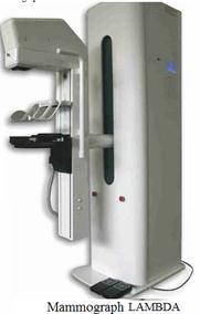
Examples of devices for x-ray diagnostics. Diagnostic medical device LAMBDA is used for mammography researches of internal structure of mammary glands. Mobile roentgen surgical device TAU9 with C-shaped arch is used in operational.


Поиск по сайту: