АвтоАвтоматизацияАрхитектураАстрономияАудитБиологияБухгалтерияВоенное делоГенетикаГеографияГеологияГосударствоДомДругоеЖурналистика и СМИИзобретательствоИностранные языкиИнформатикаИскусствоИсторияКомпьютерыКулинарияКультураЛексикологияЛитератураЛогикаМаркетингМатематикаМашиностроениеМедицинаМенеджментМеталлы и СваркаМеханикаМузыкаНаселениеОбразованиеОхрана безопасности жизниОхрана ТрудаПедагогикаПолитикаПравоПриборостроениеПрограммированиеПроизводствоПромышленностьПсихологияРадиоРегилияСвязьСоциологияСпортСтандартизацияСтроительствоТехнологииТорговляТуризмФизикаФизиологияФилософияФинансыХимияХозяйствоЦеннообразованиеЧерчениеЭкологияЭконометрикаЭкономикаЭлектроникаЮриспунденкция
Essence of a method
Electroencephalography (EEG) is a method of research of bioelectric activity a brain, arising during its activity.
The membrane of a nervous cell has the potential of rest making nearby 60-70 mkV, which is a necessary condition of normal functioning of neuron and generating of electric activity by it. At delay or the termination of a metabolism electric activity of neurons decreases, and then completely stops. It speaks about clinical and biological death of a brain.
The electric processes occurring at a level separate neuron, probably to register by means of the microelectrodes entered directly in neuron.
In a clinical practice electric activity of a brain is investigated by skin or needle electrodes. The sizes of electrodes considerably exceed of size neuron. Therefore curves of EEG is result of total electric activity of a plenty of nervous cells.
EEG of the healthy person has well organized rhythmic fluctuations that testify to presence of uniting (synchronizing) structures of a brain.
Now it is established, that regulation of functional activity of a brain is carried out, mainly, by onsetter structures. Local activization of a part of subcortical systems causes involving in process of all making active - braking structures and distribution of their influences on all brain.
 EEG is a potential difference between some point of a surface of a brain and skin electrode located behind an ear (fig. 1). The quantity of points for removal of biopotential can essentially vary (from 2 up to several decades) depending on the purposes of research.
The example of registration and kind EEG is presented on fig. 2.

 EEG looks like complex, regular fluctuation with various frequencies and amplitude. For research of electric activity of a brain at various functional conditions are usually considered spectral making (simple sine wave fluctuations of various frequencies and amplitudes on which, according to theorem Fourier, it is possible to spread out complex fluctuation -EEG).
It is very complex curve it is similar on a noise signal. During processing the received signals their Fourier-decomposition is used. For each tap spectra of dependence spectral density of capacity of signal EEG from frequency are registered.
Fig. 3.
Biopotentials of EEG and registered potential differences in 100 times are weaker, than in an electrocardiogram: 0,1 - 5 mV in an electrocardiogram and 0,001 - 0.05 mV in EEG.
Therefore for registration their strengthening in some thousand times that is reached by means of multicascade strengthening is necessary.
Electroencephalohraph «ЭНЦЕФАЛАН» allow to spend next researches.
Research of the visual and acoustical caused potentials
Research of background activity of a brain, revealing nidal changes and epileptic activity,
Three-dimensional localization of sources of electric activity of a brain,
Research visual and acoustical induced biopotential
Research of superslow activity of a brain, research of brain blood circulation.

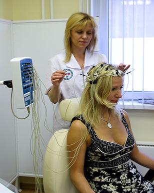
Fig. 4.
All kinds of activity of a brain in dynamics are subject to strengthening and easing, they are accompanied by the certain rhythms of electric fluctuations:
0,5-3 Hz – δ - rhythm;
4-7 Hz – θ - rhythm;
8-14 Hz – α -rhythm;
14-35 Hz – β -rhythm;
35-70 Hz – γ -rhythm.
In conditions of full rest and absence of external irritants slow rhythms of change of a condition of a brain cortex prevail. And spontaneously changing EEG it is registered - activity of a brain that finds reflection in the form of so-called an alpha - rhythm. As the basic components of spontaneous superficial EEG of the healthy person is considered with two sorts of rhythmic fluctuations of potential: α - and β - waves. An alpha - waves are characterized by frequency from 8 up to 13 impulses in sec and arise at the person at exception of visual irritant (in darkness or at the closed eyes in a condition of rest). At the majority of people α - the rhythm is well expressed. The amplitude α -waves do not exceed 50 – 100 mkV. The greatest regularity and amplitude α - rhythm is registered in parietal area of a brain cortex on border with occipital.
Transition of the person to the vigorous activity leads to change an alpha - rhythm on faster beta - rhythm. β -waves dominate in EEG of the person over an active condition, intensive physical and brainwork, an emotional pressure, realization rough and conditioned reflexes. - the rhythm consists of fast waves duration up to 40 - 50 msec and frequency 14 - 30 impulses in sec. The amplitude β -waves does not exceed 5 - 10 mkV. β - the rhythm is better shown in frontal areas of a brain cortex.
Transition from a condition of rest to a condition of the concentrated attention or to a dream is accompanied by development of slower theta - rhythm or delta - a rhythm.
The delta - a rhythm (δ -rhythm) consists of rhythmic slow waves with duration from 250 up to 1000 msec, frequency of fluctuations 1 - 4 in a second. The given rhythm is shown at a narcotic dream or at defeats of cortical departments of a brain and it has the amplitude which is not exceeding 20 - 30 mkV in EEG of the healthy person during a dream.
In EEG of the sleeping person it is possible to register and theta-rhythm (θ -rhythm) with frequency 4 - 8 fluctuations/sec. θ - the rhythm is shown and at pathological conditions of a brain, and also at an extreme emotional pressure.
When the brain gets the new fast irritation, then induced potentials of a brain (IP) are registered on EEG. They are synchronous reaction of set neurons of the given zone of a brain cortex. They consist of primary and a secondary answer on irritation that is registered in the form of positive-negative fluctuations. On EEG it is possible to analyze reaction of trained people to semantic loadings.

Fig. 5. A- the scheme of registration of EEG; B - basic rhythms of EEG.
Поиск по сайту: